Prowess Teleradiology Services
Menu
Case of the day
History
5 years male child present with history of Chronic headache and vertigo.
Description
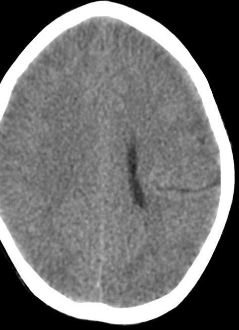
Non contrast CT Scan of the brain showed Left parietal parenchymal cleft lined by grey matter extending from the cortical surface to ipsilateral lateral ventricle.
Findings
Findings are suggestive of Schizencephaly – Closed Lip type.

Discussion
Schizencephaly
Schizencephaly is a rare congenital brain malformation characterized by clefts or deep, fluid-filled fissures in the cerebral hemispheres. It is believed to occur due to abnormal neuronal migration during fetal development. The exact etiology of schizencephaly is not known, but it has been associated with genetic mutations, infections, and environmental factors.
Radiological findings of schizencephaly include the presence of unilateral or bilateral clefts in the cerebral hemispheres, which can be visualized on magnetic resonance imaging (MRI) or computed tomography (CT) scans. The clefts are lined with gray matter and may be filled with cerebrospinal fluid. The extent and severity of the clefts can vary, and they can be classified into two types: open-lip and closed-lip schizencephaly.
Open-lip schizencephaly is characterized by a cleft that extends from the pial surface to the ventricular system, resulting in communication between the cleft and the ventricles. Closed-lip schizencephaly is characterized by a cleft that does not communicate with the ventricles.
Schizencephaly can lead to developmental delays, intellectual disability, seizures, and motor deficits. Treatment options include antiepileptic medications, physical therapy, and surgery in some cases.
In conclusion, schizencephaly is a rare congenital brain malformation with characteristic radiological findings of clefts in the cerebral hemispheres. Early diagnosis and management are crucial for improving outcomes in affected individuals.
References:
- Barkovich AJ, Kuzniecky RI, Jackson GD, Guerrini R, Dobyns WB. A developmental and genetic classification for malformations of cortical development. Neurology. 2005;65(12):1873-87.
- Barkovich AJ, Guerrini R, Kuzniecky RI, Jackson GD, Dobyns WB. A developmental and genetic classification for malformations of cortical development: update 2012. Brain. 2012;135(Pt 5):1348-69.
- Leventer RJ, Jansen A, Pilz DT, et al. Clinical and imaging heterogeneity of polymicrogyria: a study of 328 patients. Brain. 2010;133(Pt 5):1415-27.


